Fracture de la malléole latérale (fibulaire)
Définition :
Lésion fréquente
Le trait de fracture fibulaire peut-être à plusieurs niveaux :
- trait fibulaire spiroïde interligamentaire : les ligaments tibio-fibulaires inférieurs sont respectés, le diastasis est un faux diastasis intra-osseux de Vidal
- trait fibulaire siège au-dessus de la syndesmose
- au niveau de la syndesmose postérieure
- fracture de Maisonneuve
Aucune radio n'est disponible
Aucune radio n'est disponible
Vidéos
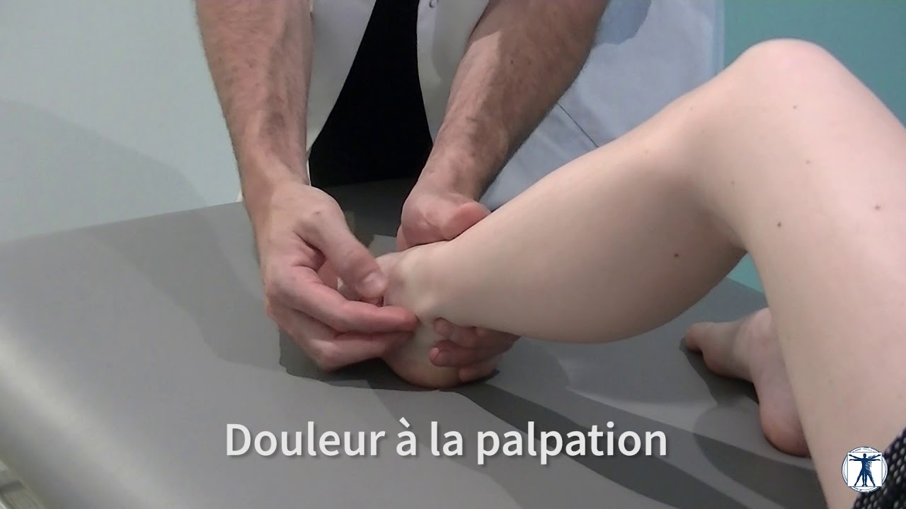
- Testing/Palping
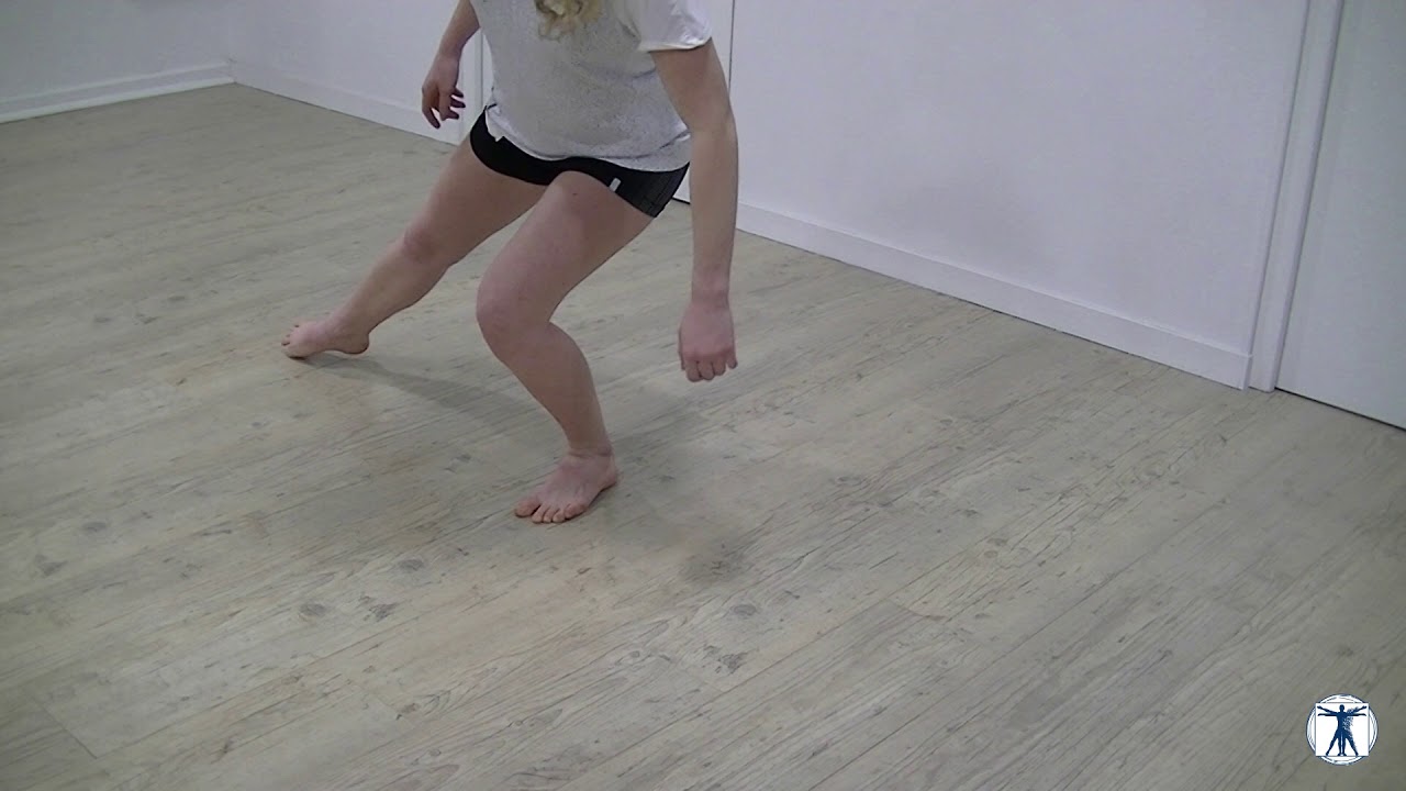
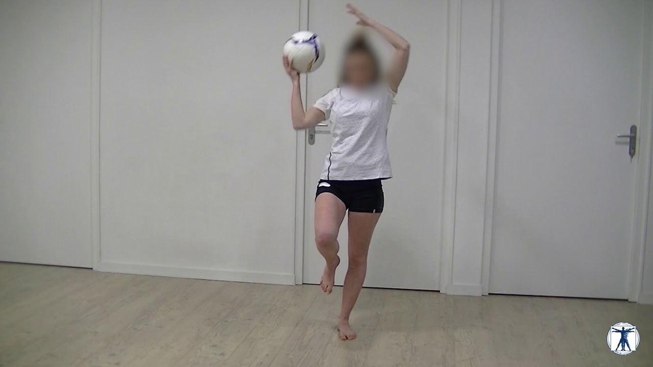
- Rééducation
Localisations :
Étiologies :
Facteurs favorisants :
Facteurs de risque :
Non renseigné
Facteurs de gravité :
Diagnostics différentiels :
Non renseigné
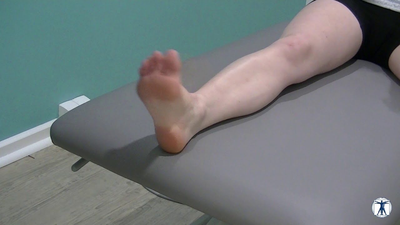
Signes cliniques :
Examens complementaires :
Orientations therapeutiques :
Traitements possibles :
Cas cliniques :
Aucun cas clinique n'a été partagé
Evaluation et préconisation
Etudes cliniques
2000 Jun;25(4):305-7.
Alkaptonuric ochronosis presenting as palmoplantar pigmentation.
We describe a 37-year-old woman who presented with palmoplantar pigmentation, thickening and pitting of 4 years duration. Bluish pigmented patches were seen over the sclera of her eyes. Her lumbar spine showed typical calcification of the intervertebral discs. Addition of Benedict's reagent to a urine sample of the patient gave rise to greenish brown precipitate and brownish black supernatant. Alkalinization of urine turned it black. A biopsy of the palmar lesion demonstrated irregular breaking up, swelling and homogenization of collagen bundles in the reticular dermis. Yellow-brown (ochre coloured) pigment was seen lying within the collagen bundles and also freely in the deeper dermis confirming our clinical diagnosis of alkaptonuric ochronosis. To the best of our knowledge this is probably the second report of alkaptonuria presenting with palmoplantar pigmentation.
Source : https://www.ncbi.nlm.nih.gov/pubmed/109714921988 Jan-Feb;16(1):60-3.
Stress fractures of the medial malleolus.
Six athletes, all engaged in running activities at the time of injury, presented with tenderness over the medial malleolus and ankle effusion. Three patients had a fracture line which could be seen on radiographs. These patients were treated by open reduction and internal fixation using two 4.0 cancellous screws. The other three patients had normal radiographs but bone scans showed increased uptake over the medial malleolus. These patients were treated with cast and immobilization. We believe that each of these patients suffered a stress fracture of the medial malleolus. We suggest that the possibility of a stress fracture be considered in the differential diagnosis of patients who present with 1) chronic or subacute pain over the medial malleolus and ankle effusion, and 2) a history of running activity at the time of injury or running activities aggravating the pain. Bone scans appear to be more sensitive than radiographs in detecting a stress fracture of the medial malleolus. We propose that athletes with radiographic signs of a medial malleolar fracture who desire early return to full participation should be treated by open reduction and internal fixation. For these patients, early motion can be initiated. Other athletes whose fracture cannot be detected on radiographs but whose malleolus shows increased uptake in the area on bone scans can be treated nonsurgically with immobilization and then progressive increase in activity. All of our patients returned to full activity between 6 and 8 weeks after treatment was initiated.
Source : https://www.ncbi.nlm.nih.gov/pubmed/33448822002 Jul;23(7):647-50.
Stress fractures of the medial malleolus--review of the literature and report of a 15-year-old elite gymnast.
Stress fracture of the medial malleolus is rare and not reported in children. We report a case of a 15-year-old elite gymnast with open physes sustaining a medial malleolar stress fracture. The patient was treated initially by rest and gradually returned to sport with full recovery. Two months later she developed a complete fracture of the medial malleolus of the same side. This was treated surgically by open reduction and internal fixation with a cancellous screw and soon after the operation she returned to full activities. Emphasis is given to the suspected mechanism which led to this unique fracture and to the hormonal aspects in the professional adolescent gymnast. We recommend surgical treatment of stress fracture of the medial malleolus especially for elite athletes, leading to early recovery and return to sports activities.
Source : https://www.ncbi.nlm.nih.gov/pubmed/121467771981 Mar;4(3):301-4. doi: 10.3928/0147-7447-19810301-08.
Isolated fracture of the lateral malleolus.
Isolated fractures of the lateral malleolus are stable only if no other ligaments are torn. These fractures are unstable if accompanied by a tear of the deltoid or anterior tibiofibular ligament and it may be necessary to demonstrate the resulting talar instability by stress radiographs. Stable fractures are best treated by the application of a short-leg walking cast. In unstable fractures an attempt should be made to anatomically reduce the lateral malleolus by closed means followed by the application of a long-leg cast. Should subsequent loss of position occur or should it not be possible to obtain an anatomical reduction, then open reduction and internal fixation is indicated.
Source : https://www.ncbi.nlm.nih.gov/pubmed/248333502011 Sep;24(9):788-90.
[Treatment of medial malleolus fractures with closed reduction and percutaneous internal fixation].
OBJECTIVE:
To study clinical effects of minimally invasive, effective and economic operational method for the treatment of medial malleolus fractures.
METHODS:
From March 2008 to August 2010, 19 patients (12 males and 7 females, ranging in age from 17 to 42 years, averaged 31.7 years) with medial malleolus fractures were reviewed. Closed reduction and percutaneous internal fixation were applied, with a hollow compression screw inserted at the centre and perpendicularly to the fracture surface. A Kirschner wire was inserted through the cortical bone of opposite side and in accordance with the axis of inner malleolus. Postoperative therapeutic effect was evaluated by Kaikkonen sprained ankle scoring system and imageology examination.
RESULTS:
All the patients got primary healing of incisions and were followed up, the duration ranged from 6 to 30 months, with an average of 18.7 months. All the patients obtained bone union. Clinical healing time ranged from 2.6 to 3.8 months, averaged 3.2 months. According to Kaikkonen scoring system, the results were rated as excellent in 5 cases, good in 10 cases, moderate in 3 cases, and poor in 1 case.
CONCLUSION:
It is a minimally invasive, effective and economic method to treat medial malleolus fractures by closed reduction and percutaneous internal fixation with hollow compression screw and Kirschner wire.
Source : https://www.ncbi.nlm.nih.gov/pubmed/22007593