Luxation du talus (astragale)
Définition :
1°degré : subluxation tibio-tarsienne
2°degré : luxation sous-astragalienne du pied
3°degré : énucléation (luxation double de l'astragale)
Aucune radio n'est disponible
Aucune radio n'est disponible
Vidéos
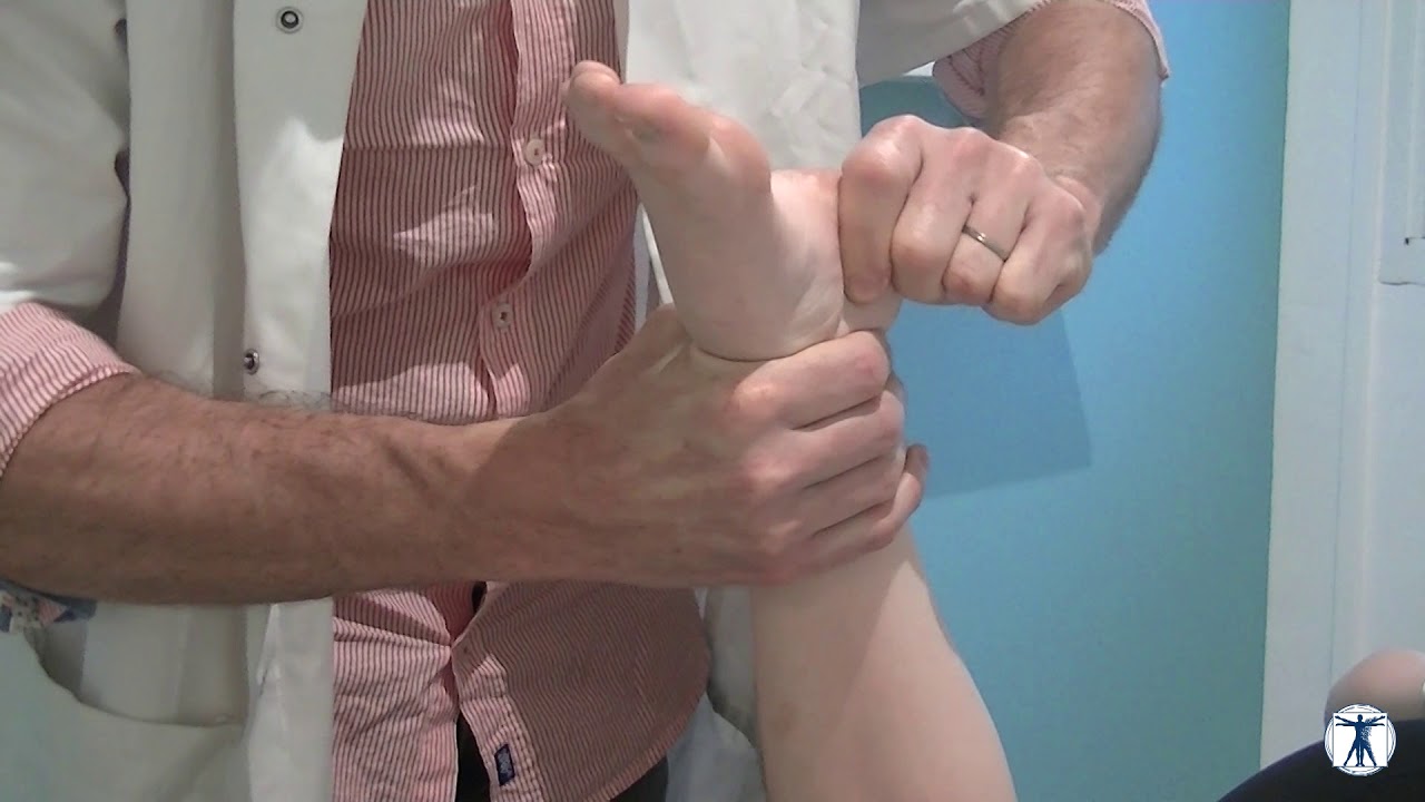
- Testing/Palping
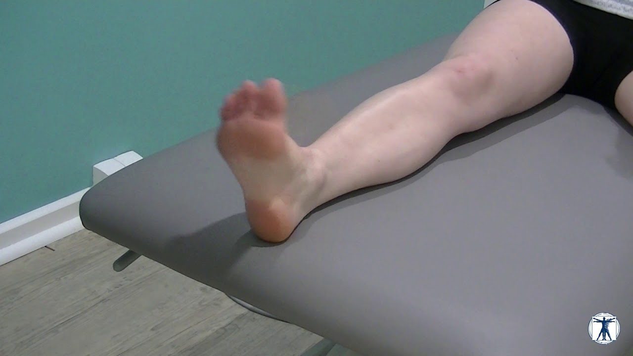
- Rééducation
Localisations :
Étiologies :
Facteurs favorisants :
Facteurs de risque :
Facteurs de gravité :
Diagnostics différentiels :
Non renseigné
Signes cliniques :
Examens complementaires :
Orientations therapeutiques :
Traitements possibles :
Cas cliniques :
Aucun cas clinique n'a été partagé
Evaluation et préconisation
Etudes cliniques
2000 Jun;25(4):305-7.
Alkaptonuric ochronosis presenting as palmoplantar pigmentation.
We describe a 37-year-old woman who presented with palmoplantar pigmentation, thickening and pitting of 4 years duration. Bluish pigmented patches were seen over the sclera of her eyes. Her lumbar spine showed typical calcification of the intervertebral discs. Addition of Benedict's reagent to a urine sample of the patient gave rise to greenish brown precipitate and brownish black supernatant. Alkalinization of urine turned it black. A biopsy of the palmar lesion demonstrated irregular breaking up, swelling and homogenization of collagen bundles in the reticular dermis. Yellow-brown (ochre coloured) pigment was seen lying within the collagen bundles and also freely in the deeper dermis confirming our clinical diagnosis of alkaptonuric ochronosis. To the best of our knowledge this is probably the second report of alkaptonuria presenting with palmoplantar pigmentation.
Source : https://www.ncbi.nlm.nih.gov/pubmed/109714922013;62:79-91.
Fractures and dislocations of the midfoot: Lisfranc and Chopart injuries.
The midfoot is a complex association of five bones and many articulations between the forefoot metatarsals and the talus and calcaneus, which make up the hindfoot. These anatomic relationships are connected and restrained by an even more complex network of ligaments, capsules, and fascia, which must function as a unit to provide normal and painless locomotion. The common eponyms of Lisfranc and Chopart refer to the distal and proximal joint relationships of the midfoot, respectively. Midfoot injuries range from single ligament strains to complicated fracture-dislocations involving multiple bones and joints. To provide best outcomes for patients, it is important to understand the anatomy and the mechanical function of the midfoot; to review the epidemiology, mechanism, and classification of injuries encountered in an orthopaedic clinical practice; and to review the principles, indications, and surgical techniques for managing midfoot fractures and dislocations.
Source : https://www.ncbi.nlm.nih.gov/pubmed/233950162012 Jul-Aug;26(4):235-44.
[Long-term results of the treatment of Lisfranc fracture dislocation].
We conducted an ambispective cohort study of 83 patients with a diagnosis of Lisfranc fracture dislocation from 1993 to 2008. The lesions were classified into two groups: pure dislocations and fracture dislocations of the Lisfranc joint using the Hardcastle-Reschamer classification. The results included the following data: sociodemographic and epidemiologic variables, lesion-related variables, clinical parameters, and the following clinical and functional assessment scales: Baltimore Painful Foot Score, Creighton-Nebraska Health Foundation, American Orthopaedic Foot and Ankle Society (AOFAS), and Hannover Scoring System. Sixty-three patients were treated surgically. Closed reduction and minimally invasive fixation with Kirschner nails were performed in 53 patients (63.9%), and open reduction with a dorsal approach and fixation with Kirschner nails in 10 cases (15.2%). In 46 cases de medial column was fixed, in 61 cases the intermediate column, and in 42 the lateral column. Sixty-six (79.5%) of the patients had complications including both acute and late ones. Regardless of the technique used, the purpose of treatment was the anatomical reduction of the involved joints. Based on our experience, we think that the use of Kirschner nails is effective, as it provides enough stiffness and stability. In general terms, this injury is not as disabling as it had been considered in the literature. Patients consider their discomfort as tolerable and compatible with the demands of their activities of daily living and they may perform their work considering the time limitations.
Source : https://www.ncbi.nlm.nih.gov/pubmed/233203262011 Mar 23.
Epidemiology, imaging, and treatment of Lisfranc fracture-dislocations revisited.
The purpose of this article is to discuss the features of Lisfranc injuries and identify their typical imaging findings on radiographs, CT, and MR imaging. Lisfranc injuries are most often caused by hyperplantarflexion of the foot, often during a sporting injury or in high-speed motor vehicle collisions. The most common radiographic findings include diastasis of the base of the first and second metatarsals and the "fleck" sign, though neither is necessarily present in every Lisfranc fracture-dislocation. Owing to their often subtle radiographic presentation, clinically suspected Lisfranc injuries warrant imaging with a more sensitive test for the detection of osseous and ligamentous Lisfranc injuries. 3D CT imaging provides a comprehensive evaluation of the injury for optimal treatment planning, with resultant decreased long-term patient morbidity. Furthermore, 3D volume-rendered CT and CT multiplanar reconstructions (MPRs) provide osseous and neurovascular anatomic detail that may be a considerable help with surgical planning for operative cases of Lisfranc injuries. Also, with 3D CT and MPRs, other occult fractures, which are common in patients with high-energy injury and multiple trauma, may become evident.
Source : https://www.ncbi.nlm.nih.gov/pubmed/214314382014 Oct 9.
Staged treatment of high energy midfoot fracture dislocations.
BACKGROUND:
Staged care with interval external fixation is a successful established treatment strategy for high energy periarticular fractures with often extensive soft tissue damage such as the tibial plateau and plafond. The aim of the current study was to determine whether staged care of high energy midfoot fracture/dislocation with interval external fixation prior to definitive open reconstruction in the polytraumatized patient was both safe and efficacious.
METHODS:
One hundred twenty-three patients were operated on for high energy midfoot fracture/dislocation during the 8-year study period. Eighteen polytrauma patients were selectively treated with a staged protocol. Radiographic assessment was utilized to determine if the fixator achieved gross skeletal alignment. Further, final alignment after definitive reconstruction and postoperative complications were analyzed.
RESULTS:
The fixator improved both length and alignment of all high energy midfoot fracture/dislocations. Loss of acceptable reduction while in the temporary frame occurred in only 1 case. Final alignment after definitive reconstruction was anatomic in all cases. No cases of wound-related complication and/or deep infection occurred.
CONCLUSION:
Delayed reconstruction of high energy midfoot fracture/dislocation using interval external fixation should be an accepted care paradigm in selected polytrauma patients.
LEVEL OF EVIDENCE:
Level III, retrospective comparative study.
Source : https://www.ncbi.nlm.nih.gov/pubmed/253018902012 Jul-Sep;116(3):834-9.
Lisfranc midfoot dislocations: correlations between surgical treatment and functional outcomes.
Lisfranc dislocations and fracture dislocations are the most common severe injuries of the foot.
AIM:
To assess the functional outcome of patients with Lisfranc dislocations of the midfoot by applying the latest methods of diagnosis and treatment.
MATERIAL AND METHODS:
The study reviewed 31 patients with dislocations and fracture- dislocations of the Lisfranc joint over a 10 years period. The average follow-up period was 44 months (range 12-108). Injuries were classified according to Myerson scale.
RESULTS:
The outcomes were evaluated using the Baltimore Painful Foot score (PFS) and American Orthopaedic Foot and Ankle Society (AOFAS) mid-foot scoring scale. 10 patients had an excellent outcome on the PFS scale, 8 were classified as good, 13 fair and poor. The average AOFAS score for the midfoot. used for results interpretation was 72 (range 52-92).
CONCLUSIONS:
Of all methods of surgical treatment used, the highest scores were obtained by internal fixation with screws. Eight patients (25.8 %) developed posttraumatic arthritis of the tarsometatarsal joints.
Source : https://www.ncbi.nlm.nih.gov/pubmed/232725382010 Dec;18(12):718-28.
Treatment of Lisfranc joint injury: current concepts.
Injuries to the tarsometatarsal joint complex, also known as the Lisfranc joint, are relatively uncommon. However, the importance of an accurate diagnosis cannot be overstated. These injuries, especially when missed, may result in considerable long-term disability as the result of posttraumatic arthritis. A high level of suspicion, recognition of the clinical signs of injury, and appropriate radiographic studies are needed for correct diagnosis. When surgery is indicated, closed reduction with percutaneous screw fixation should be attempted. If reduction is questionable, open reduction should be performed. Screw fixation remains the traditional fixation technique.
Source : https://www.ncbi.nlm.nih.gov/pubmed/211191381997 Oct;100(10):787-91.
[Mobile flatfoot as a sequela of dislocation injury of the Lisfranc joint. A retrospective analysis of 13 patients].
In this retrospective study, 13 patients with subtle Lisfranc joint injuries were examined after a mean period of 23 months using clinical assessment, radiography and dynamic pedographic gait analysis. The aims were to identify the factors leading to a mobile flatfoot deformity, evaluate the functional and clinical outcome of these injuries, and draw practical conclusions for initial management and subsequent intervention. All patients showed a mobile flatfoot deformity, increased motion in the subtalar joint, increased load on the hindfoot, decreased load on the forefoot, and a prolonged contact phase during the stance phase. Radiographs revealed progressive osteoarthrosis in the joint and a residual displacement of the medial Lisfranc joint. An unstable medial Lisfranc joint results in the development of a mobile flatfoot. Initial treatment of a subtly displaced Lisfranc joint should consist of exact anatomical reduction and additional maintenance of the longitudinal arch of the foot. After failed initial treatment, early arthrodesis of the midfoot is recommended as a salvage procedure for the foot.
Source : https://www.ncbi.nlm.nih.gov/pubmed/94462331991;84(3):159-64.
[The treatment of fresh Lisfranc dislocations and fracture-dislocations].
Dislocations and fracture dislocations of the tarsometatarsal joint are usually the result of a high energy trauma to the forefoot. A missed diagnosis or an insufficient treatment or a massive destruction of the tarsometatarsal joint result in a high rate of late morbidity. An appropriate radiological assessment, open anatomical reduction and temporary K-wire arthrodesis followed by a functional after-treatment can improve the long-term results of this severe forefoot injuries. The long term results (7 months to 20 years) of 24 tarsometatarsal injuries are analysed and a concept of their treatment presented.
Source : https://www.ncbi.nlm.nih.gov/pubmed/17602382004 May;33(3):362-4.
Plantar dislocation of lateral tarsometatarsal joint: a case of subtle Lisfranc injury.
NTRODUCTION:
We present a rare case of plantar dislocation of the cuboid, fourth and fifth metatarsal joints. Fracture-dislocation in the midfoot region may be subtle and difficult to recognise at the emergency department.
CLINICAL PICTURE:
A 16-year-old girl presented with lateral foot pain and swelling following a fall from a height of 3 m. Initial radiograph revealed a third metatarsal shaft fracture; however, additional views reviewed a plantar direction of fourth and fifth metatarsal dislocation from the cuboid.
TREATMENT:
Open reduction and internal fixation with Kirschner wires followed by immobilisation in plaster was performed.
OUTCOME:
She made an uneventful recovery and the wires had since been removed.
CONCLUSION:
This case was unusual in that there was lateral tarsometatarsal disruption with neither diastasis between the first and second metatarsals nor injury to either the first or second tarsometatarsal joints. The plantar direction of dislocation was also unusual as these injuries are usually dorsal.
Source : https://www.ncbi.nlm.nih.gov/pubmed/151757811998 Nov-Dec;55(6):665-8.
[Dislocation of the Lisfranc joint in a military skier].
The case of Lisfranc's dislocation in soldiers-skiers has been presented. The aim of the study is to realize the role of skiing equipment in generation of the injuries in soldiers-skiers. Early therapeutic intervention provides good results.
Source : https://www.ncbi.nlm.nih.gov/pubmed/100633912003 Mar 20.
Concomitant plantar tarsometatarsal (Lisfranc) and metatarsophalangeal joint dislocations.
We report an unusual case of concomitant plantar tarsometatarsal (Lisfranc) and 1st and 2nd metatarsophalangeal (MTP) joint dislocations and fracture of the neck of the third metatarsal bone which has never been reported before. The plantar dislocation of the Lisfranc joint was treated by open reduction and fixation with K-wires; the dislocations of the MTP joints and neck fracture of the third metatarsal bone were treated by closed reduction and percutaneous fixation with K-wires and immobilized with a plaster cast. At the 5 year follow-up examination, our patient had no complaints, but the radiograph showed degenerative changes of the Lisfranc and the 1st MTP joint
Source : https://www.ncbi.nlm.nih.gov/pubmed/12721687Dislocation, Lisfranc.
A Lisfranc dislocation or injury typically describes a spectrum of injuries involving the tarsometatarsal joints of the foot. The Lisfranc joint itself is composed of the articulation between the first, second, and third metatarsals bones, and the cuneiform bones. Injuries of the joint can range from complete tarsometatarsal displacement with associated fractures and ligamentous tears to partial sprains with no displacement. Although a Lisfranc injury can involve various parts of the foot the Lisfranc ligament itself is an isolated ligament that connects the medial cuneiform to the second metatarsal. Lisfranc joint injuries are uncommon and are often misdiagnosed and mismanaged. It is important to recognize these injuries early and start treatment promptly because failure to recognize and treat these will lead to midfoot arthritis, chronic pain, and functional instability. Even when recognized and treated promptly, there is still a high risk for chronic disability and complications.
Source : https://www.ncbi.nlm.nih.gov/pubmed/288463061996 Apr;25(4):305-9.
Lisfranc dislocation and associated metatarsophalangeal joint dislocations. A case report and literature review.
A case report of a 28-year-old man with associated dislocations of the tarsometatarsal (Lisfranc) and metatarsophalangeal joints is presented. The potential for disability after these injuries is very high when the diagnosis or treatment is delayed, the reduction is incomplete, or the dislocation recurs. Relevant aspects of diagnosis and treatment are discussed.
Source : https://www.ncbi.nlm.nih.gov/pubmed/87283682001 Jan;19(1):71-5.
Orthopedic pitfalls in the ED: Lisfranc fracture-dislocation.
Lisfranc fracture-dislocation of the foot is an injury that carries a high incidence of chronic pain and disability. Its emergency department presentation can be subtle, and more frequent than previously believed. This review article examines the clinical presentation, historical factors, diagnostic techniques, and management options applicable to the emergency practitioner.
Source : https://www.ncbi.nlm.nih.gov/pubmed/111460251989 Mar;92(3):130-9.
[Dislocation fractures of the Chopart and Lisfranc joint].
Dislocation fractures of Chopart's and Lisfranc's (mediotarsal) articulation result from the effects of impact and, because there are many possible complications, require a high level of experience on the part of the treating traumatologist. To avoid residual joint incongruities and intra-articular osteochondral fragments with subsequent early arthrosis and the corresponding complaints, the indications for open reduction and functionally stable osteosynthesis should be broad. The osteosynthesis can be achieved by transfixation with adjustable screws and 3.5 or 2.7 mm screws suitable for small fragments. This permits postoperative treatment to be carried out with a walking cast rather than a full plaster cast. In emergency patients with multiple injuries, dislocation fractures in the tarsal region require immediate and definitive treatment in order to avoid grave consequences and pain severe enough to influence the quality of life.
Source : https://www.ncbi.nlm.nih.gov/pubmed/27111971991 Apr;83(4):366-9.
Lisfranc fracture-dislocations: report of two cases.
Lisfranc fracture-dislocation is a rare but sometimes dramatic injury, most commonly associated with motor vehicle and industrial accidents, and falls from heights. Radiographic examination is a crucial point in the work-up of these patients. The most common findings on plain radiographs are separation of the bases of the first and second metatarsals and the "fleck" sign, a bony fragment resulting from a fracture of the base of the first or second metatarsal. These fractures are demonstrated in the two cases presented. We conclude that these lesions can usually be diagnosed with plain radiographs if scrupulous attention is paid when evaluating the normal anatomic alignments. Computed tomography should be reserved for equivocal cases.
Source : https://www.ncbi.nlm.nih.gov/pubmed/19205111985 Oct;122(10):585-6.
[Original treatment of luxation of Lisfranc's joint].
Emergency reduction and stabilization of a case of total external luxation of Lisfranc's joint with fracture of 3rd cuneiform was by partial internal arthrodesis using a 1/3 tube plate placed astride the tarso-metatarsal joint combined with plaster immobilization for 2 months. Recovery took 6 months.
Source : 1989;43(7):562-7.
[Fracture-luxation of the Lisfranc joint. Apropos of 39 cases].
Lisfranc fracture-dislocations are relatively rare (two per thousand). They generally occur in multiple trauma victims and may pass unnoticed. Open reduction followed by pinned osteosynthesis and plaster immobilisation ensures a good anatomical and functional result.
Source : https://www.ncbi.nlm.nih.gov/pubmed/2619223